AFM-based characterization of peptide arrays
Allergies, ebola or cancer – many diseases that affect millions have unclear etymology and challenging diagnostic procedures. Multiple diagnostic tests can already be done in parallel using a lab-on-thechip approach: Realized with high density dots (peptide arrays) on a glass slide by the group of PD Dr. Alexander Nesterov-Müller. However, so far the only way to see the disease-causing peptides was by detection of a fluorescent signal coming from died antibodies. Thus, important quantitative information about peptide arrays like their morphology, roughness and density were missing. That is where Dr. Julia Syurik‘s expertise in characterizing morphology of polymeric surfaces with atomic force microscope (AFM) comes in. The idea came up at a YIN-get-together and was realized due to the YIN-Grant.
The project profits from the synergies of both groups and develops new insights into peptide synthesis surfaces with nanoscale characterization of their morphology and phase contrast. The change of
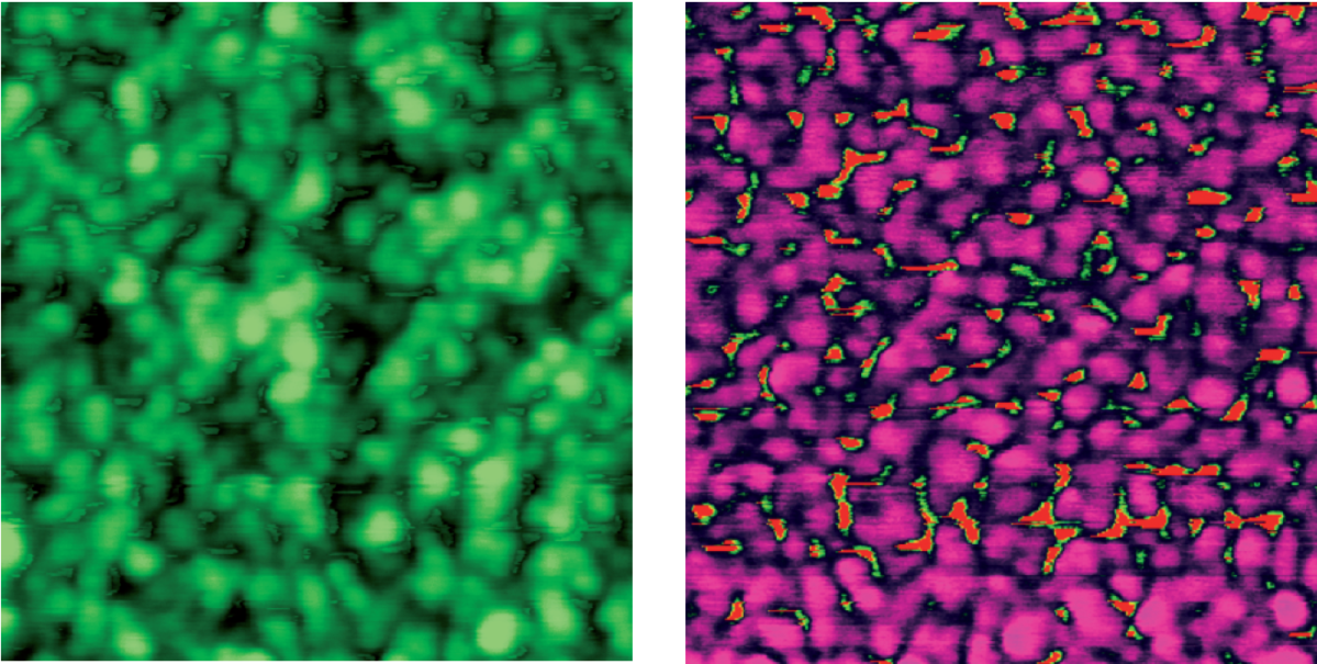
morphology for a full creation process of a peptide array was measured and analyzed. The pictures show AFM images of a PEG polymer layer after solid phase peptide synthesis and staining with antibodies. As PEG is a polymer blend, it is not surprising to see phase segregation on the morphology image (left). However, to see the strong inhomogeneity on the phase image (right) was unexpected. A 63 % phase difference suggests that peptides attach to the valleys in the PEG matrix, which was not known before. The minimal size of the islands with a nonpolymer material is 8 nm, what corresponds to the radius of the cantilever.
These results can be used to understand protein folding, protein-protein interactions in vivo, cell migration and for development of novel bio inspired electronic materials. The novel methodology allows comprehensive varying of peptides in high density array format, thus giving clues for protein manipulation.

